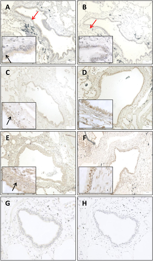
Figure 2.
Representative images of LXRα and LXRβ expression in small airway epithelium and subepithelium. The distribution of LXRα (A, C, and E) and LXRβ (B, D, and F) in the epithelium (red arrow in A) and subepithelium (red arrow in B) of small airways, present in lung sections of non-smoking controls (NS) (A-B), smoking controls (S) (C-D) and COPD patients (E-F). LXRα and LXRβ were detected using 3,3’-diaminobenzidine (brown; positive LXRα stain indicated by black arrow) and cell nuclei were counterstained using Meyer’s haematoxylin. Substitution of LXRα (G) and LXRβ (H) primary antibodies for isotype controls displayed no immunoreactivity.

