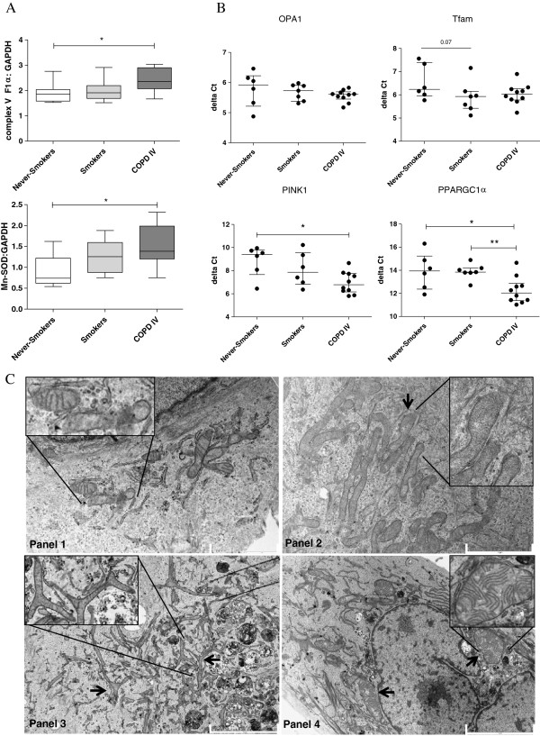
Figure 5.
Primary bronchial epithelial cells (PBECs) from COPD patients display elongated and swollen mitochondria, fragmentation, branching and cristae depletion and altered expression of mitochondrial markers compared to control PBECs. PBECs were isolated from ex-smoking COPD patients with GOLD stage IV (n = 10), control smokers (n = 7) and never-smokers (n = 6) A) Complex V ATPase protein and Mn-SOD were detected by western blotting. GAPDH was used as loading control. B) OPA1, Tfam, PINK1 and PPARGC1α mRNA expression was detected by qPCR and related to housekeeping genes. ΔCt values are shown and median interquartile ranges (IQR) are indicated. C) Panel 1 shows a representative picture of PBECs from 4 never-smokers displaying normal mitochondria. Panel 2 shows a representative picture from 9 ex-smoking COPD patients displaying increased numbers of mitochondria with cristae depletion and ’club shaped’ ends. Panel 3 COPD patient shows severe branching. Panel 4 COPD patient shows swollen structures. Scale bar indicates 5 μm (bottom) and 2 μm (Top). p = 0.07, * = p < 0.05 and ** = p < 0.01 between the indicated values.

