
Figure 10.
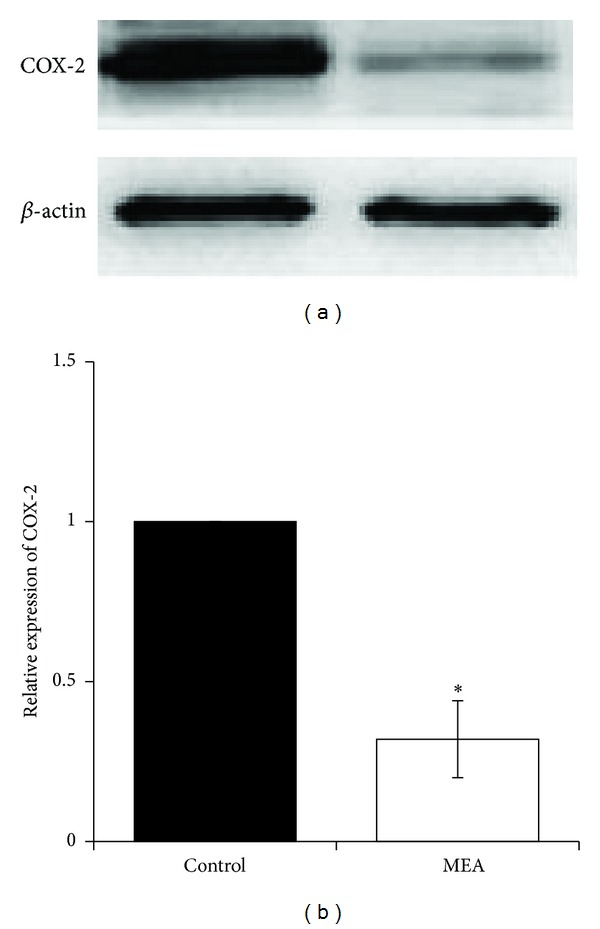
Protein expression of COX-2 24 hours after transient MCAO in different groups. (a) Representative immunoblots of COX-2 in the right cerebral hemisphere of different groups 24 hours after transient MCAO. (b) Semiquantitative analysis of protein expression of COX-2. Data are expressed as means ± SEM (n = 3). *P < 0.01, compared with the control group. The relative expression of COX-2 was significantly decreased by MEA pretreatment.