Figure 8.

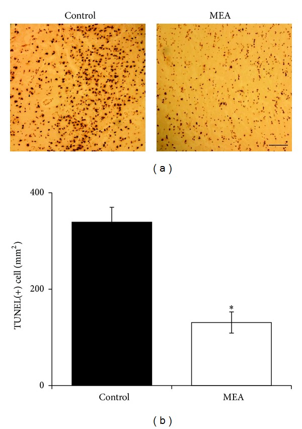
Apoptosis in the penumbra of right ischemic cerebral hemisphere 24 hours after transient MCAO. (a) Representative images of TUNEL staining in the penumbra of right ischemic cerebral hemisphere 24 hours after transient MCAO. The brown staining within the nuclei reveals TUNEL-positive cells. (b) Quantitative analysis of TUNEL-positive cells. Data are expressed as means ± SEM (n = 5). *P < 0.01, compared with the control group. The number of TUNEL-positive cells was significantly decreased by MEA pretreatment. Scale bar = 100 μm.
