
Figure 1.
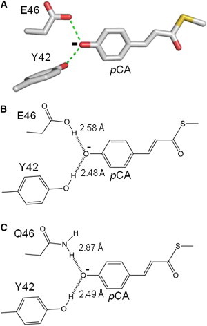
Anionic hydrogen bonding of pCA at the active site of PYP. A three-dimensional depiction of hydrogen bonding of the anionic pCA at the active site of wtPYP based on its crystal structure (PDB ID 1NWZ) (A). A two-dimensional depiction of hydrogen bonding of the pCA with residues 42 and 46, including hydrogen bonding lengths based on the crystal structures of wtPYP (B) and E46Q PYP (C) (PDB IDs: 1NWZ and 1OTA). To see this figure in color, go online.