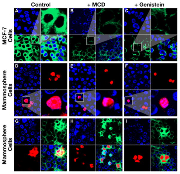
Figure 5.
BCS cells have a significantly higher rate of caveolin-independent endocytosis than the differentiated breast cancer cells. A. Staining of the differentiated breast cancer MCF-7 cells with LacCer in the absence of CVDE inhibitor: top left, cells were stained with DAPI; bottom left, with BODIPY-labeled LacCer; top right, amplified image of the cells from bottom left section; bottom right, merge of the top left section and bottom left section. B. Staining of the differentiated breast cancer MCF-7 cells with LacCer in the presence of CVDE inhibitor MCD. C. Staining of the differentiated breast cancer MCF-7 cells with LacCer in the presence of CVDE inhibitor genistein. D. Staining of the mammosphere cells derived from MCF-7 with BCS/A35 in the absence of CVDE inhibitor: top left, cells were stained with DAPI; top right, with TR-labeled BCS/A35; bottom left, merge of the top left section and top right section; bottom right, amplified image of the cells from bottom left section. E. Staining of the cells derived from mammospheres with BCS/A35 in the presence of CVDE inhibitor MCD. F. Staining of the cells derived from mammospheres with BCS/A35 in the presence of CVDE inhibitor genistein. G. Co-staining of the cells derived from mammospheres with BCS/A35 and LacCer in the absence of CVDE inhibitor: top left, cells were stained with DAPI; top right, with BODIPY-labeled LacCer; bottom left, with TR-labeled BCS/A35; bottom right, merge of the previous three sections. H. Co-staining of the cells derived from mammospheres with BCS/A35 and LacCer in the presence of CVDE inhibitor MCD. I. Co-staining of the cells derived from mammospheres with BCS/A35 and LacCer in the presence of CVDE inhibitor genistein.

