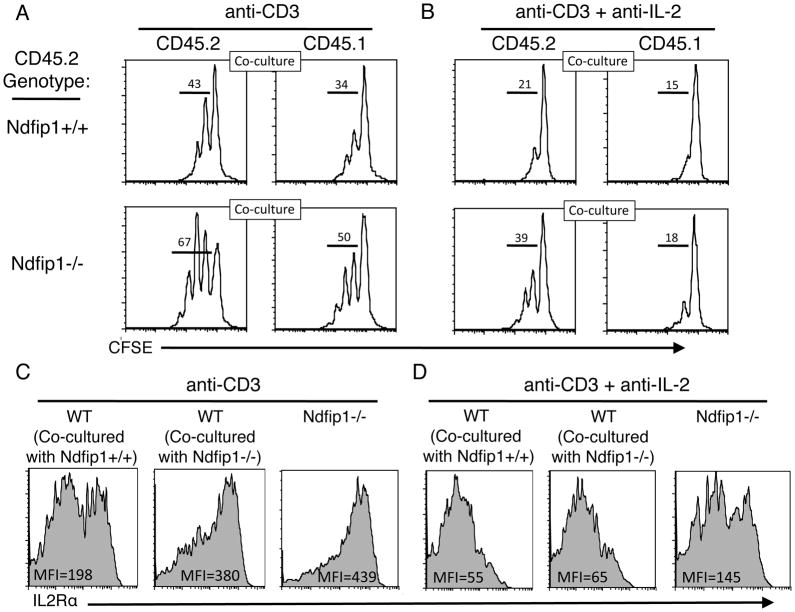
Figure 6. IL-2 production by Ndfip1−/− T cells can increase the IL-2Rα levels and allow proliferation of WT T cells.
(A–D) Naïve Ndfip1+/+ or Ndfip1−/− T cells (both are CD45.2) were mixed with equal numbers of WT (CD45.1) T cells, labeled with CFSE, and cultured in the presence of anti-CD3. Data are representative of two independent experiments, which together represent 4 mice from each genotype. (A) After 3 days, cells were analyzed for proliferation as determined by CFSE dilution and results are shown in histograms. (B) Anti-IL-2 was added to the cultures at day 0 and cells were again analyzed at day three for proliferation as described for panel A. (C) IL-2Rα levels were analyzed on the cells in panel A. Mean fluorescence intensities (MFI) for the population are shown within each histogram. (D) IL-2Rα levels were analyzed on the cells in panel B. The MFI for the population are shown within each histogram.

