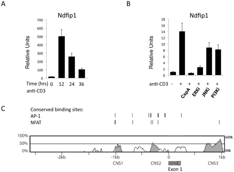
Figure 8. Inhibitors for NFAT and Erk prevent the expression of Ndfip1 in TCR-stimulated cells.
(A–B) Naïve CD4+ T cells were stimulated with anti-CD3 and Ndfip1 mRNA levels were measured at the indicated time points (A) or after overnight stimulation (B). (B) Cells were cultured in the presence of the indicated inhibitors. Cyclosporine A (cspA) inhibits NFAT. (A–B) mRNA levels were normalized to an internal control and data shown are relative to unstimulated levels. Bars represent the mean and lines show standard deviation of triplicate samples. Data is representative of 3 different experiments using three or more mice per genotype. (C) Comparison of conserved non-coding sequences in the Ndfip1 promoter between mouse and man. The red and green box between CNS2 and CNS3 indicates the signal peptide and exon 1 (respectively) of the Ndfip1 gene.

