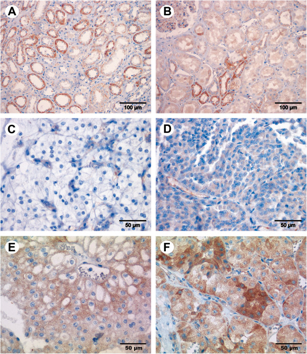
Figure 1.
Immunostaining of RECK in renal cell tissue. RECK expression was mainly seen in the tubuli and in the capillaries of the glomeruli shown here in the medulla of non-malignant kidney tissue (A) and in the non-malignant renal cortex (B). RECK expression increased from clear cell carcinoma (C) over papillary (D) and chromophobe (E) carcinoma to oncocytoma (F). Magnification: 200× (A, B), 400× (C-F).

