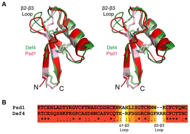
Figure 6. Structural homology between MtDef4 and other plant defensins.
A. Shown here is the structural alignment of MtDef4 and the plant defensin Psd1 from Pisum sativum (PDB code 1JKZ) in green and red, respectively. The cysteine residues involved in the four disulfide bonds are highlighted in lighter hues.
B. Sequence alignments based on the 3D alignments using the program EXPRESSO [27]. The coloring of the alignments ranges from blue to red as the error in the alignment goes from high to low. Note that the β2-β3 loop in MtDef4 is longer by two residues and is far more basic than that in Psd1. However, the exposed hydrophobic F37 residue in MtDef4 is highly similar to W38 residue in Psd1.

