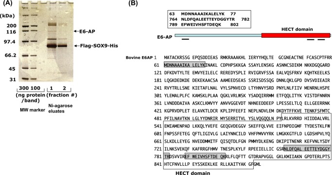
FIGURE 1.
Proteomics analysis of FLAG-SOX9-His-binding nuclear proteins. A, after nuclear extracts from fetal bovine rib chondrocytes expressing FLAG- and His-tagged SOX9 were loaded onto nickel-agarose (Ni-agarose), the eluates were further purified on anti-FLAG-agarose. Bound proteins were precipitated with 10% TCA, resolved by SDS gel electrophoresis on 10% BisTris gel, and stained with silver. For the control, the same amount of nuclear extract was prepared from non-transfected chondrocytes, but no significant bands were detected (not shown). All SOX9-binding bands were excised from the gel individually, and eluted proteins were digested with trypsin and subjected to MS/MS analysis. MW, molecular weight. B, three peptides that were sequenced corresponded to the ubiquitin ligase E6-AP. The locations of these peptides are shown as gray shadows in the entire amino acid sequence of E6-AP. One peptide is located in the N terminus, and the other two are in the HECT domain.

