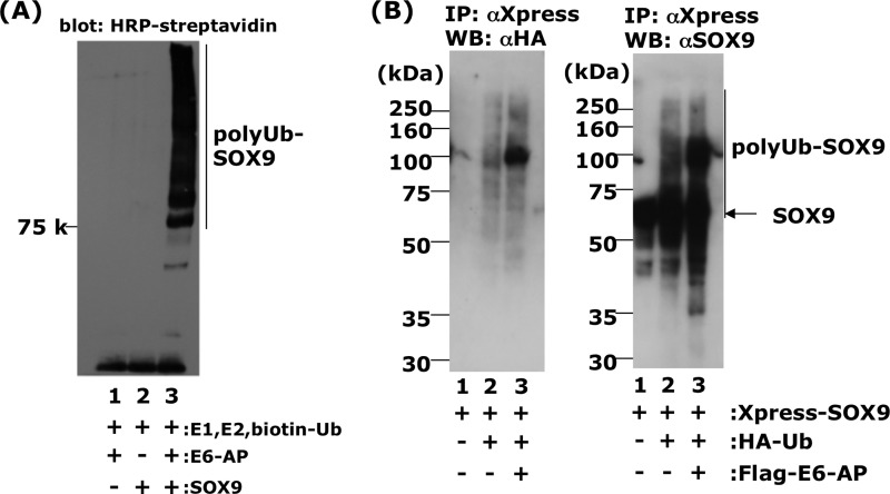
FIGURE 5.
E6-AP enhances SOX9 ubiquitination in vivo and in vitro. A, ubiquitination of recombinant SOX9 in vitro in the presence of E1, E2 (UbcH5c), biotinylated ubiquitin, ATP, and E6-AP (lane 3). In lane 2, E6-AP was omitted from the reaction mixture. In lane 1, SOX9 was omitted from the reaction mixture. B, ubiquitination of SOX9 in vivo. HEK293 cells were cotransfected with 0.5 μg of pcDNA4/HisMax-SOX9 (lane 1), 0.5 μg of pcDNA4/HisMax-SOX9, plus 1 μg of pcDNA3.1-HA-ubiquitin (HA-Ub) (lane 2), 0.5 μg of pcDNA4/HisMax-SOX9, 1 μg of pcDNA3.1-HA-ubiquitin, and 4 μg of pFLAG-E6-AP (lane 3). Twenty-four hours after transfection, the cells were treated with 2 μm MG132, a proteasome inhibitor, and further incubated for 15 h. After 15 h, the cells were disrupted by sonication, immunoprecipitated (IP) with anti-Xpress antibody to precipitate Xpress-SOX9 complexes, and Western blotted (WB) with anti-HA antibody for detection of ubiquitinated protein (B, left panel) and anti-SOX9 antibody (B, right panel) as described under “Experimental Procedures.” In lane 3, some anti-SOX9-positive bands (polyUb-Sox9) were migrating more slowly than the major SOX9 band.

