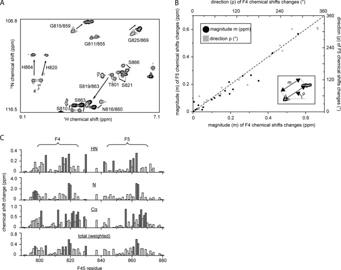
FIGURE 2.
Both F4 and F5 bind DNA using the same subset of residues. A, portion of the 15N HSQC of 15N-labeled double ZF construct F4F5 in the absence (gray) and presence (black) of 1 molar eq of RARE DNA. Assignments are indicated. B, graph showing magnitude (black dots, left and bottom axes) and direction (gray boxes, right and top axes) of chemical shift changes (measured from 15N HSQC spectra as indicated in the inset) that occur for residues in F4 as compared with the corresponding residues in F5 upon the addition of 1 molar eq of RARE DNA. Only residues 802–826 in F4 and 846–870 in F5 are shown. The high correlation between F4 and F5 changes for a given residue strongly suggests the same mode of binding for both ZFs. C, summary of chemical shift changes for HN, N, and Cα nuclei. Total chemical shift changes, weighted according to Ayed et al. (36), are also shown.

