
FIGURE 2.
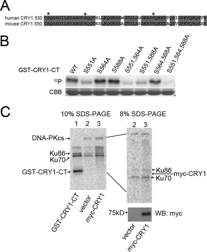
DNA-PK can phosphorylate isolated CRY1 C-terminal tails but not the intact CRY1 protein. A, sequence alignment of human (top row) and mouse (bottom row) CRY1 C-terminal tails. Asterisks mark the three serines in SQ motifs. Gray shading denotes conserved amino acids. B, in vitro DNA-PK kinase assays using isolated CRY1 C-terminal tails (WT and various mutants, as noted) as substrates. 32P shows phosphorylation. CBB is Coomassie Brilliant Blue staining of total GST-CRY1-CT showing equal loading in each lane. C, in vitro DNA-PK kinase assay on full-length CRY1 protein. The left panel shows autoradiography of a 10% polyacrylamide gel, which shows autophosphorylated DNA-PKcs (lanes 2 and 3), phosphorylated Ku86 and Ku70 (components of the DNA-PK complex that are also autophosphorylated), and phosphorylation of the C-terminal tail (lane 1). However, no phosphorylation of full-length CRY1 is observed. The right panel is an expanded view of the same samples as lanes 2 and 3 of the left panel, run on 8% gel to better separate the region where CRY1 would migrate (expected CRY1 position marked by an arrow). The bottom right panel is a Western blot (WB) of the samples in lanes 2 and 3, demonstrating that sample 3 does contain mCRY1 protein.