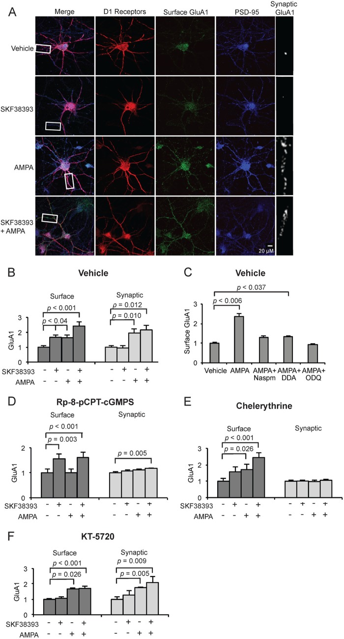
FIGURE 3.
CPARs selectively induce GluA1 trafficking through GluA1 Ser-845 phosphorylation. A, visualization of surface and synaptic GluA1 in cultured MSNs expressing D1 receptors (10 DIV) after treatment with AMPA (5 μm, 1 min) and/or SKF38393 (10 μm, 5 min) and 30 min pretreatment with APV (10 μm), CdCl2 (50 μm), TTX (500 nm). Neurons were stained for D1R, surface GluA1, and PSD-95 (synaptic marker). Scale bar = 20 μm. B, quantification of surface and synaptic GluA1 in cultured MSNs treated in A; n = 25 dendrite regions. C, MSNs (10 DIV) were treated with vehicle or AMPA (50 μm, 1 min) either alone or in cultures pretreated for 30 min with NASPM (200 μm), 2,3-dideoxyadenosine (DDA; 10 μm), or 1H-[1,2,4]oxadiazolo[4,3-a]quinoxalin-1-one or ODQ (5 μm), and surface GluA1 levels were determined and normalized to vehicle; n = 20 dendrite regions. D–F, MSN cultures were treated as in A and pretreated either with Rp-8-pCPT-cGMPS (D), chelerythrine (E), or KT-5720 (F). Data are represented as mean pixel intensity ± S.E. normalized to vehicle treatment and analyzed using one-way ANOVA followed by Fisher's post hoc tests; n = 20 dendrite regions for each condition.

