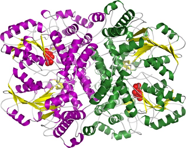
FIGURE 3.

Overall structure of the Synechocystis P-protein homodimer in the reduced state with the cofactor PLP bound. Shown is a ribbon diagram with β-sheets in yellow and helices in purple in one subunit and green in the second subunit. PLP is shown as red spheres.
