
FIGURE 1.
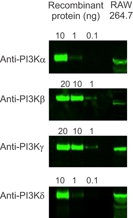
PI3K isoform expression in RAW264.7 cells. First to third lanes illustrate different amounts of recombinant PI3K isoforms, as indicated at the top. Lysates from undifferentiated RAW264.7 cells are shown in the fourth lane, immunoblotted with isoform-specific anti-PI3K antibodies, with evidence for all four isoforms. Immunoblot data are representative of two independent cell preparations.