
FIGURE 4.
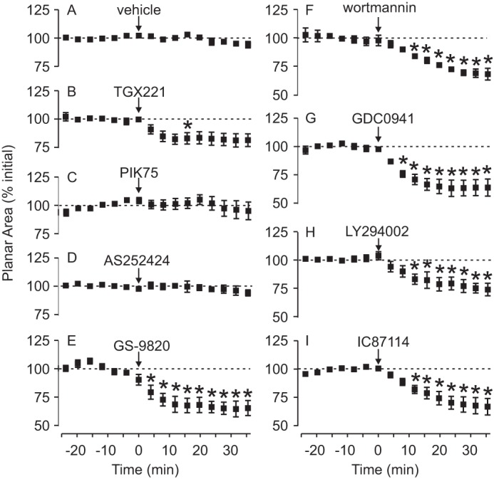
PI3Kδ is important in osteoclast lamellipodia spreading. Rat osteoclasts were imaged by time-lapse phase-contrast microscopy and treated with the indicated test substance at time 0, as described in the legend to Fig. 3. Image analysis software was used to calculate the planar area of osteoclasts at 4-min intervals. Data are expressed as a percentage of the mean initial area before time 0 (from −24 to 0 min) and are mean ± S.E. A, there was no marked change in osteoclast area in vehicle-treated cells (n = 4 independent preparations, a total of 9 osteoclasts). B–D, the following inhibitors had no consistent effect on osteoclast area: TGX221 (1 μm, n = 8 independent preparations, a total of 10 osteoclasts), PIK75 (1 μm, n = 5 independent preparations, a total of 10 osteoclasts), and AS252424 (1 μm, n = 5 independent preparations, a total of 9 osteoclasts). E–I, in contrast, the following inhibitors caused significant, sustained decreases in osteoclast planar area: GS-9820 (1 μm, n = 5 independent preparations, a total of 15 osteoclasts), wortmannin (1 μm, n = 4 independent preparations, a total of 8 osteoclasts), GDC0941 (1 μm, n = 5 independent preparations, a total of 8 osteoclasts), LY294002 (50 μm, n = 3 independent preparations, a total of 7 osteoclasts), and IC87114 (5 μm, n = 4 independent preparations, a total of 12 osteoclasts). Differences were assessed using a two-way ANOVA and Bonferroni's multiple comparisons test. * indicates p < 0.05 compared with vehicle at the corresponding times.