
FIGURE 7.
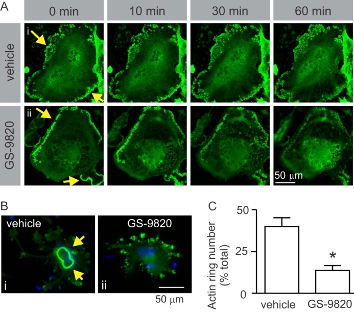
Inhibition of PI3Kδ causes disruption of actin cytoskeleton. A, live-cell imaging of the effects of GS-9820 on F-actin belt dynamics in rabbit osteoclast expressing actin-EGFP. To directly observe the effects of GS-9820 on F-actin belts, rabbit osteoclasts were plated on FBS-coated MatTek glass-bottom culture dishes and transduced with adenoviruses expressing actin-EGFP fusion or EGFP proteins. Cells were then bathed in HEPES-buffered M199 medium with 15% FBS at ∼26 °C and imaged using confocal microscopy (×40 objective, Zeiss LSM 510 META confocal microscope). Images labeled 0 min illustrate the appearance of osteoclasts immediately prior to addition of vehicle or GS-9820 (1 μm) to the bath. Yellow arrows indicate actin belt. A, i, F-actin belt remained intact in vehicle-treated osteoclast. A, ii, GS-9820 induced gradual disappearance of the peripheral actin structures and an increase in actin fluorescence more centrally. Images are representative of a total of 4 osteoclasts from 2 independent preparations. Control samples transduced with EGFP protein alone showed uniform distribution of fluorescence (not shown). B, to examine effects of PI3K on sealing zone formation, rat osteoclasts were plated on resorbable calcium phosphate-coated discs. Samples were treated with vehicle or GS-9820 (1 μm) for 10 min and then fixed. F-actin was labeled using Alexa Fluor 488-conjugated phalloidin (green), nuclei were stained with DAPI (blue), and cells were examined by fluorescence microscopy. B, i, vehicle-treated osteoclast exhibits a prominent F-actin-rich sealing zone, characteristic of active osteoclasts on resorbable substrates. Note that sealing zones are more centrally located than the peripheral actin belts illustrated in A. B, ii, osteoclasts treated with GS-9820 exhibited fewer sealing zones and disorganized clusters of F-actin. C, osteoclasts treated with GS-9820 displayed significantly fewer sealing zones (actin rings) than vehicle-treated cells. Data are the percentage of osteoclasts exhibiting sealing zones and are mean ± S.E., n = 3 independent experiments with a total of 592 osteoclasts. Data were analyzed by unpaired Student's t test. * indicates p < 0.05 compared with vehicle.