FIGURE 6.
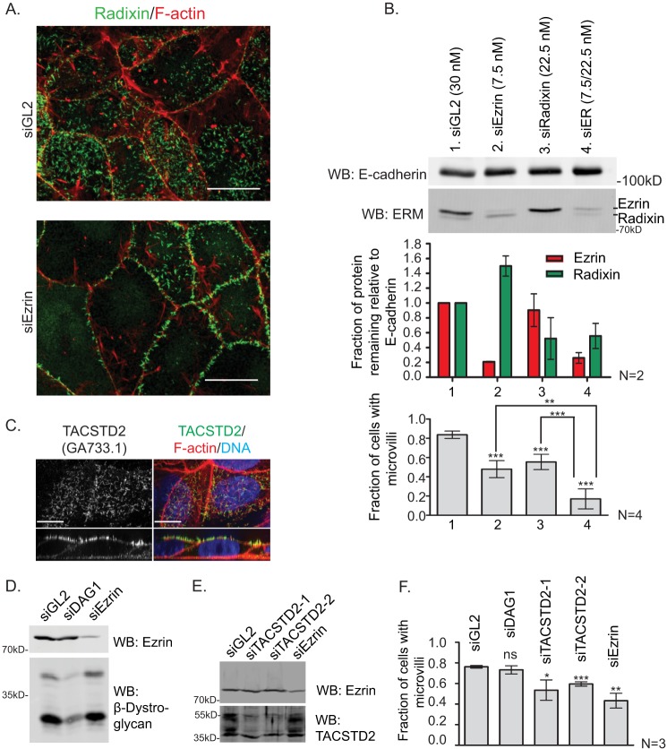
TACSTD2 suppression by RNA interference results in a moderate loss-of-microvilli phenotype. A, Jeg-3 cells were transfected with nontargeting control siRNA (siGL2) or ezrin siRNA (siEzrin) for 72 h and then stained for F-actin and radixin to reveal a loss of microvilli after to ezrin knockdown. Scale bars, 10 μm. B, Jeg-3 cells were transfected with indicated siRNAs for 72 h, and cell lysates were prepared and Western-blotted (WB) for ezrin and radixin. The remaining protein was quantified. The cells were also fixed and stained for F-actin, β-dystroglycan, and EBP50 (data not shown). The presence of microvilli by any of these markers was scored using a previously described scheme (7, 15, 17, 18, 49). Error bars are standard deviation. p values were computed using a two-tailed t test. *, p < 0.05; **, p < 0.01; ***, p < 0.001; ns, not significant. C, endogenous TACSTD2 was detected by immunofluorescence using the GA733.1 antibody. Scale bars, 10 μm. D and E, cells were transfected with indicated siRNA for 72 h, then lysates were prepared and Western-blotted (WB) as indicated. F, Jeg-3 cells transfected as described in B were stained for radixin, and the presence of microvilli was scored as in B. Error bars are standard deviation. p values were computed using a two-tailed t test. *, p < 0.05; **, p < 0.01, ***; p < 0.001; ns, not significant.

