FIGURE 10.
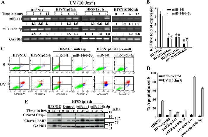
p16-CDK4-Sp1-dependent up-regulation of miR-141 and miR-146b-5p and their role in apoptosis following UV damage. A, cells were either mock-treated or challenged with UV light (10 Jm−2) and then re-incubated for the indicated periods of time. Total RNA was then isolated and used for RT-PCR using specific primers for the indicated genes. The numbers below the bands indicate the corresponding expression levels following loading correction against GAPDH. B, qRT-PCR on mRNAs corresponding to 2 h of post-irradiation relative to time 0. These experiments were performed in triplicate (*, p < 0.05). C, cells were either mock-treated or challenged with UV light (10 Jm−2) and incubated for 72 h. Apoptosis was analyzed by annexin V/propidium iodide flow cytometry. D, the histogram shows the proportions of spontaneous and induced apoptosis (early + late). The error bars represent standard deviations of at least three different experiments. E, HFSN1C and HFSN1p16sh cells expressing the control plasmid, pre-miR-141 or pre-miR-146b-5p, were treated as above and collected after 48 and 72 h. Whole cell lysates were prepared and used for immunoblotting analysis. Error bars represent means ± S.D.

