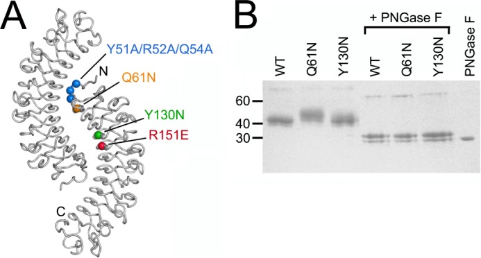
FIGURE 1.

Mouse decorin mutants. A, location of mutations in mouse decorin mapped onto the crystal structure of the bovine decorin dimer (14). The dimer is viewed along its symmetry axis, and the N and C termini are labeled in one subunit. B, reducing SDS-PAGE of wild-type (WT) mouse decorin and the Q61N and Y130N mutants before and after digestion with peptide N-glycosidase F (PNGase F) (Coomassie Blue stain). The positions of selected molecular mass markers are indicated on the left.
