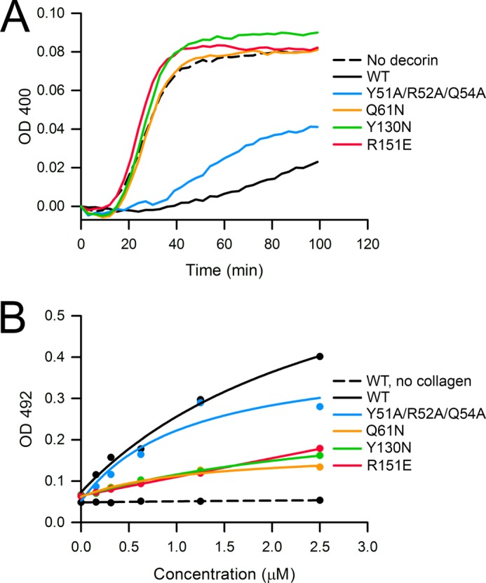
FIGURE 6.

Collagen binding by WT and mutant mouse decorin. A, inhibition of collagen fibrillogenesis by WT and mutant mouse decorin. Type I collagen (32 μg/ml) was incubated at pH 7.8 and 37 °C, and the turbidity arising from fibril formation was recorded as absorbance at 400 nm. The decorin proteins were added at a concentration of 50 μg/ml. Shown is a representative of three independent experiments. B, collagen binding by WT and mutant mouse decorin. Type I collagen was immobilized on microtiter plates and incubated with varying amounts of decorin proteins. Bound decorin proteins were detected as absorbance at 492 nm using an antibody-linked color reaction. The solid lines are fits of the data by an equation describing single site binding. Shown is a representative of three independent experiments carried out in duplicate.
