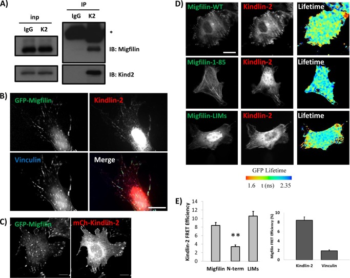
FIGURE 4.
Migfilin co-localizes and exhibits specific FRET with kindlin-2 in cells. A, endogenous migfilin and kindlin-2 co-immunoprecipitate from NIH3T3 lysates. * = antibody heavy chain. B, GFP-migfilin co-localizes with endogenous kindlin-2 and vinculin in NIH3T3 fibroblasts plated on fibronectin. Scale bar = 20 μm. C, GFP-migfilin co-localizes with mCherry-kindlin-2 in NIH3T3 fibroblasts plated on fibronectin. Scale bar = 10 μm. D, fibroblasts on fibronectin coexpress GFP-migfilin constructs and mCherry-kindlin-2. Individual channels are shown and lifetime of the donor fluorophore is shown in pseudocolor, where cyan indicates less FRET and red/yellow indicates more FRET (see bar). Scale bar, 20 μm. E, quantification of kindlin-migfilin or migfilin-vinculin FRET efficiency. **, p value < 0.001.

