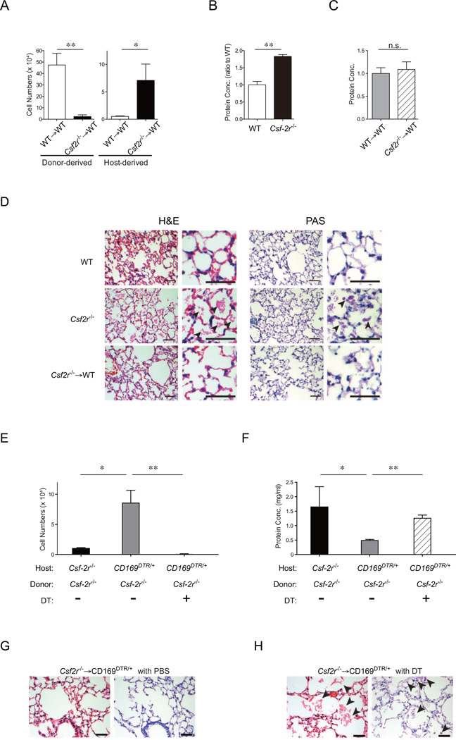
Figure 6. Lung macrophages repopulate locally after lethal irradiation and promote lung tissue integrity.
(A-D) CD45.1+ mice were lethally irradiated and transplanted with 5 × 10⁶ BM cells isolated from CD45.2+ Csf-2r−/− (black bars, n=6) mice or control CD45.2+ littermate (white bars, n=5). (A) The absolute numbers of donor- and host-derived lung macrophages in right lung lobes were enumerated 2 months after bone marrow transplantation. (B, C) Protein concentrations in the bronchial alveolar lavage fluid (BALF) were quantified in wild-type mice (white bar), Csf-2r−/− mice (black bar), and wild-type mice transplanted with wild-type (gray bar) or Csf-2r−/− (hatched bar) BM (n=3/group) two months after bone marrow transplantation. Data were pooled from two independent experiments and shown as mean +/− SEM. (D) Representative sections of left lung lobes stained with H&E (left) and PAS (right) obtained from steady-state wild-type (WT) mice (top), steady-state Csf-2r−/− mice (center), and WT mice transplanted with Csf-2r−/− (bottom) BM. Arrow heads: PAS+ eosinophilic material within the alveolar spaces. Scale bar: 25µm. (E-H) Csf-2r−/− mice and CD169DTR/+ mice were lethally irradiated and reconstituted with 5 × 10⁶ BM cells isolated from C57BL/6 WT or Csf-2r−/− mice. A group of CD169DTR/+Csf-2r+/+ recipients were injected with DT (10µg/kg, twice weekly) starting from day +3 post-transplant. The absolute numbers of macrophages in the right lung lobes (E) and protein concentrations in BALF (F) in [Csf-2r−/− into Csf-2r−/−] (black bars, n=3) and [Csf-2r−/− into CD169DTR/+] chimera treated with PBS (gray bars, n=6) or DT (hatched bars, n=6) were enumerated two months after transplant. Data were pooled from two independent experiments and shown as mean +/− SEM. Paraffin sections of the left lung lobes of recipients of [Csf-2r−/− into CD169DTR/+] chimeras treated with PBS (G) or DT (H) at 2 months post transplant were stained with H&E (left panel) and PAS stain (right panel). Sections isolated from [Csf-2r−/− into CD169DTR/+] chimera treated with DT (H) demonstrate granular eosinophilic material positive for PAS within the alveolar spaces (arrow heads) whereas sections from [Csf-2r−/− into CD169DTR/+] chimera treated with diluent (G) demonstrate normal alveolar structure. Scale bars: 25 µm. See also Figure S6. *: P < 0.05; **: P < 0.01.

