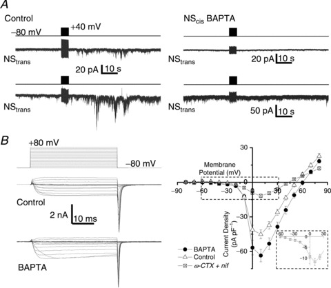
Figure 1. Block of SS transmission by a rapid Ca2+ scavenger.

A, two paired NS were patch clamped simultaneously and both were held at −80 mV. A stimulus train (2 ms pulse to +40 mV at 100 Hz for 5 s, as indicated by upper protocol trace for each trial) was delivered to one neuron, designated NScis (not shown but as in Rozanski et al. 2012) with (right panel) or without (left panel) intracellular BAPTA while recording from its passive neuron pair, NStrans. B, Ca2+ current traces recorded from NScis evoked by a family of depolarizing voltage pulses, as indicated in the left upper panel, in the absence (left middle panel) and presence (left lower panel) of intracellular BAPTA (10 mm). The right panel plots steady state (mean amplitude of the last approximately 10 ms of the depolarizing pulse) current amplitudes against the pulse voltage for control (n= 14) and BAPTA (n= 15) neurons. The mean current-to-voltage relationship is also shown for NScis neurons in the presence of nifedipine (nif) and ω-conotoxin GIVA (ωCTX; both 2 μm, n= 11; current traces are shown in Fig. 2A). The inset is an expanded view of the boxed region and shows an LVA current shoulder.
