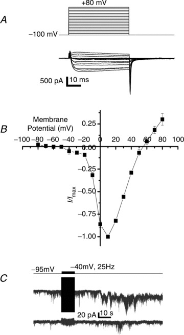
Figure 5. Role of LVA channels in SS transmission.

A, current traces (bottom panel) evoked in NScis by depolarizing step potentials (top panel) in the presence of nifedipine (2 μm) and ω-conotoxin GIVA (2 μm). B, current to voltage plot for n= 4 recordings as in A. C, trace recorded from NStrans held at −80 mV during a 25 Hz, −40 mV amplitude and 20 ms duration pulse train delivered to NScis (upper protocol trace).
