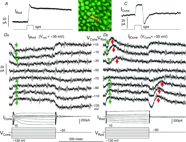
Figure 1. Synaptic interactions between a rod and a next-neighboring cone.

A and C, simultaneous light-evoked current responses of the rod and the cone to a 500 nm, −3.0, 0.5-s light step; B, Lucifer Yellow fluorescent image of the rod and cone outer segments in the retinal flatmount. Calibration bar: 20 μm. Da, current responses of the rod (IRod, held near its dark potential at −35 mV) to voltage clamp steps in the cone (VCone) from −50 mV to various voltages (−130 to +10 mV, middle column in D), ICone traces in Da are current responses of the cone to the voltage steps in itself. Db, current responses of the cone (ICone, held near its dark potential at −35 mV) to the same set of voltage clamp steps in the rod (VRod), IRod traces in Db are current responses of the rod to the voltage steps in itself. Green arrows: sustained, bi-directional transjunctional currents (Ij); red arrows: transient rod→cone currents (IRC).
