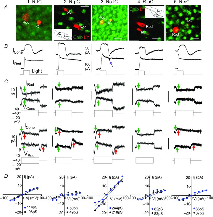
Figure 2. Synaptic interactions between rods and cones.

Lucifer Yellow-filled cell images in flat-mounted salamander retinas (A, calibration bars: 20 μm), simultaneous light-evoked current responses (B), bi-directional current responses of follower cells to positive and negative voltage clamp steps to driver cells (C), and Ij–V relations (D, blue, rod→cone; black, cone→rod) of (from left to right) a rod–large single cone (R–lC) pair, a rod–principal member of a double cone (R–pC) pair, a rodC–large single cone (RC–lC) pair, a rod–accessory member of a double cone (R–aC) pair and a rod–small single cone (R–sC) pair. Green arrows in C mark the sign-preserving, bi-directional sustained Ij, and red arrows mark the sign-inverting, rod→cone transient currents IRC in cones at positive voltage step onsets and negative voltage step offsets in rods. The purple arrow in B points to the transient OFF current notch characteristic of rodC light response. Red labels in A are Lucifer Yellow (LY) images of the rod–cone pairs, and green labels in the third and fifth panels are Calbindin-immunoreactive aCs in dark fields (photoreceptors were slightly flattened for better viewing of the pCs and aCs whose contours are shown in the insets), whereas green labels in other panels are recoverin staining of cones (heavier) and rods. The slopes (Gj) of each Ij–Vj relation in panel D are given in picosiemens (pS) at lower right corner of each plot.
