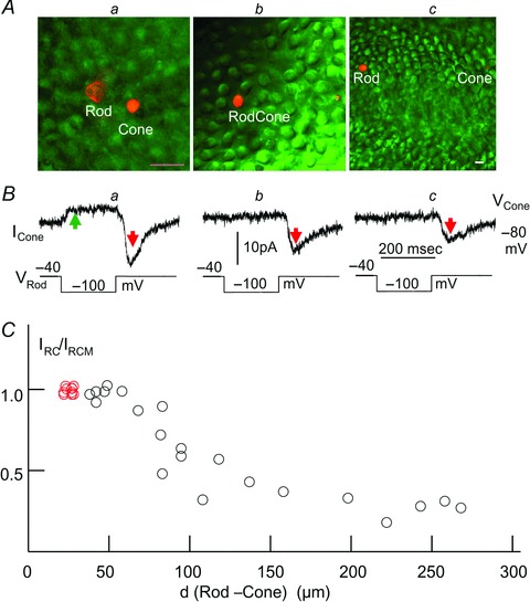
Figure 4. IRC between rods and cones of different separations.

A, Lucifer Yellow fluorescent image of the rod and cone outer segments (calibration bars: 20 μm) separated by about 20 μm (a), 95 μm (b) and 260 μm (c) in the retinal flatmounts. B, current responses of the cones held at −80 mV to a negative voltage step (−40 to −100 mV) in the rods (same 3 rod–cone pairs in Aa–c). The rod voltage step elicited both the sustained, sign-preserving Ij (green arrow) and the transient, sign-inverting IRC (red arrows), shown in a, and elicited IRC of smaller amplitude but no Ij, shown b and c. C, scatter plot of the normalized IRC–distance (IRC/IRCMvs. d (rod–cone)) relation for 15 next-neighbouring rod–cone pairs (with IRCM, red circles) and 21 rod–cone pairs (18 R–lCs, 2 R–pCs and 1 RC–lC) separated by various distances.
