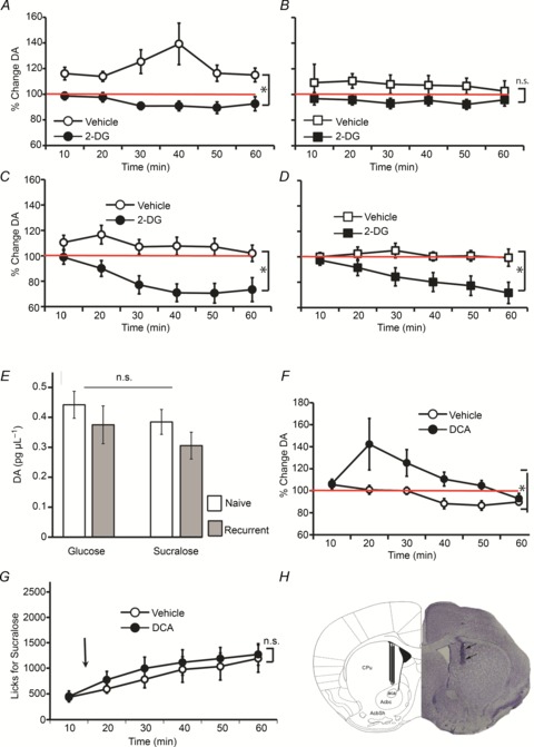
Figure 2. Dopamine efflux in mediodorsal striatum reflects the effects of glucoprivation on sugar and artificial sweetener intake.

During the intake sessions depicted in Fig. 1C–F, microdialysis sampling from the mediodorsal striatum was performed concomitantly to intake monitoring. Baseline sampling was performed for ∼30 min prior to introduction of stimulus sippers to the cages. A, striatal dopamine levels were significantly higher than baseline levels during glucose intake after vehicle injections, whereas during glucoprivation levels were not significantly different from baseline (glucoprivation effect *P < 0.001). B, in recurrent mice, striatal dopamine levels were not significantly different from baseline levels during glucose intake irrespective of the glucoprivic treatment. C, striatal dopamine levels were not significantly different from baseline levels during sucralose intake after vehicle injections, whereas these levels were significantly lower than baseline during glucoprivation (glucoprivation effect *P= 0.002). D, similar effects were observed in recurrent animals ingesting sucralose (glucoprivation effect *P < 0.05). E, baseline dopamine levels did not differ across groups. F, mice (n= 6) ingesting sucralose following an injection of dichloroacetate (‘DCA’), which promotes glucose metabolism, displayed dopamine efflux significantly above baseline levels in contrast to following vehicle injections (treatment effect *P= 0.03). G, as expected, no significant behavioural modifications were observed in response to the dichloroacetate treatment. H, Histological analyses of brain tissue obtained from the animals that performed the 1 h behavioural + microdialysis tests reveal that dopamine sampling was restricted to the most medial aspect of the dorsal striatum region. A representative case is shown on the right side of the figure along with schematic representation of the final locations overlaid on the corresponding stereotaxic map. aca, Anterior commissure; AcbC, core region of the nucleus accumbens; AcbSh, shell region of the nucleus accumbens; CPu, caudate/putamen.
