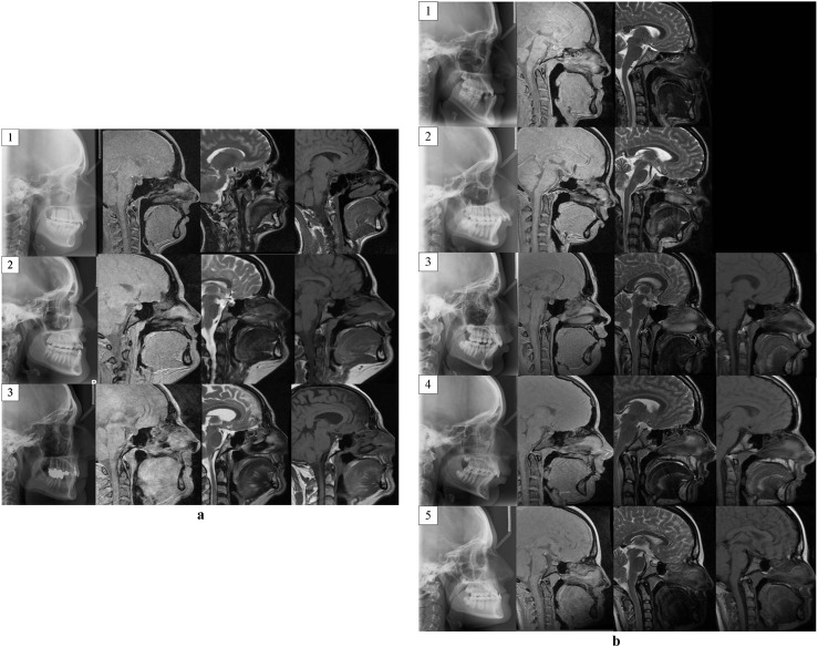
Figure 1.
(a) Lateral cephalogram (left), sagittal “Black Bone” (second from left), sagittal T2 weighted (second from right) and sagittal T1 weighted MRI images of the three volunteers (indicated by numbers 1–3) used for cephalometric analysis. (b) Lateral cephalogram (left), sagittal “Black Bone” (second from left), sagittal T2 weighted (second from right) and sagittal T1 weighted MRI images of the five patients (indicated by numbers 1–5) included for cephalometric analysis. Sagittal T1 imaging was not acquired in Patients 1 and 2

