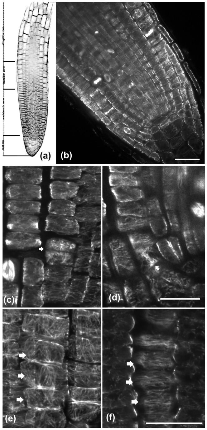
Figure 1. CLSM images of wild-type A. thaliana roots after staining with FM4-64 (a) or after tubulin immunostaining (b-f).
(a) CLSM longitudinal section through the root tip, illustrating the apical root zones. (b) Cortical microtubules are predominantly transverse in interphase cells of the meristematic zone. (c) In cells that have just divided (arrow) microtubules are randomly oriented. (d) The arrow points a cell close to the quiescent center, dividing almost parallel to the root axis. (e, f) Microtubule organization in protodermal cells of the meristematic zone (marked by arrows). (e) Maximum projection of CLSM sections of the external periclinal cell face. Cortical microtubules exhibit a loose longitudinal orientation. (f) CLSM section through the inner periclinal cell face, where cortical microtubules are transverse. The root tip in these images as well as in the following ones is located towards the bottom of the page. Scale bars, 20 μm.

