
Figure 5.
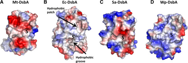
Electrostatic molecular surface of Mt-DsbA compared to other DsbA homologs. A. Mt-DsbA (PDB ID: 4IHU) reveals strong negative charges around the hydrophobic active site. B. Ec-DsbA, PDB ID: 1DSB, [40]C. Sa-DsbA, PDB ID: 3BCI, [31]D. Wp-DsbA, PDB ID: 3F4T, [42]. Positive and negative electrostatic potentials, shown in blue (+3.0 kT/e) and red (-3.0 kT/e), respectively. S represents the active sites containing the conserved CXXC motifs.