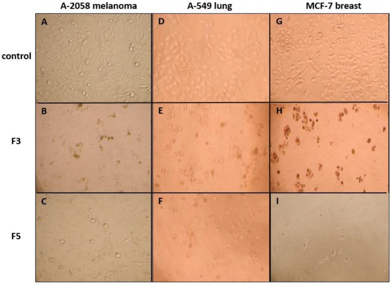
Figure 2.
Morphological changes in A-2058 melanoma cells, A-549 lung carcinoma cells and MCF-7 breast adenocarcinoma cells after a 72 h exposition to a control cell culture medium (A, D, G) or to a medium containing 100 µg·mL−1 of F3 (B, E, H) or F5 (C, F, I). Condensation and fragmentation into apoptotic bodies unequivocally demonstrated the strong cytotoxicity of F3 and F5 and suggested their pro-apoptotic effect.

