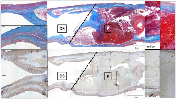
Figure 1.

Longitudinal section of an undivided CEA specimen at artery center line level showing the cutting line for P and DS segments separation. Prevalent complicated plaque features of P (lipid-necrotic core, calcium deposits, fibrosis and haemorrage) and milder changes of its downstream side DS (prevalent VSMCs and collagen component, small lipid and calcium deposits) are evidenced by Masson’s trichrome and α-SMA immunostaining (from top to bottom). Original magnification 2×, insets 10×.
