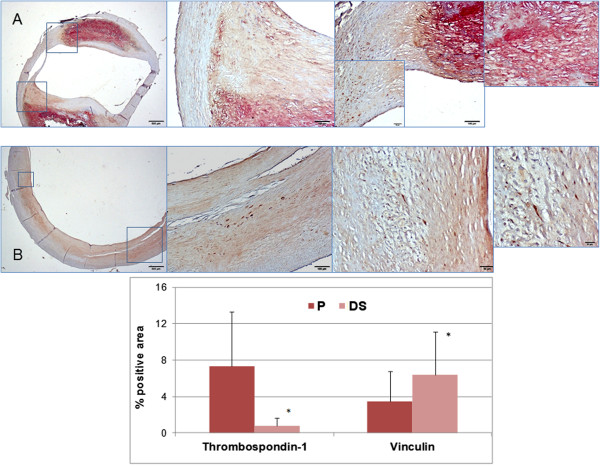
Figure 6.

Top: Double immunostaining of thrombospondin-1 (red) and vinculin (brown) in P (A) and DS (B) sections of a fibrocalcific type Vb plaque. Low magnification (2×, left) and high magnification (10× to 40×) microscopic fields (insets) are shown. Neither cellular nor extracellular co-distribution of the two antibodies is present. A selective cellular binding of vinculin both on P an DS sections is evident, while thrombospondin-1 is almost exclusively located in the fibrocalcific core of P. Bottom: Average values and SD of thrombospondin-1 and vinculin positivity (single immunostaining) in lesional area of all CEA specimens showing a markedly greater stain in P as compared toDS for thrombospondin-1 and an opposite pattern for vinculin. *p<0.02 paired t-test.
