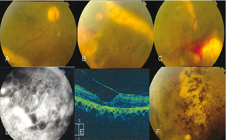
Figure 2.
Retinitis pigmentosa (RP) associated with vasoproliferative tumor (VPT) and Coats like fundus in both eyes of case 2. A, Hazy fundus photograph of the right eye shows a dilated vein and far peripheral hard exudates inferotemporally. B, Peripheral view of the fundus in the right eye shows hard exudate precipitations around the VPT along the stuffed venules by hard exudates and telangiectatic vessels on VPT with bony spicular changes. C, VPT with telangiectatic vessels and hard exudates, note preretinal hemorrhages on and around the lesion in the left eye. D, Peripheral lesion with fluorescence surrounding the capillary nonperfusion; note light bulbs and aneurysmal telangiectatic vessels in the right eye. E, Epiretinal membrane with antero-posterior traction in OCT (OD). F, Regressed inferotemporal lesion with consequent gliosis and epiretinal membranous changes (OD).

