
Figure 5.
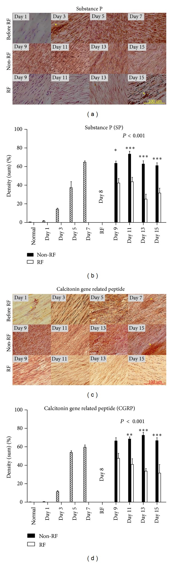
SP and CGRP stained brown in high cellularity areas. RF-treated tissues stained less SP and CGRP than tendons without RF ((a) and (b)). SP and CGRP expression were significantly reduced in RF-treated tendons ((c) and (d), n = 5, P < 0.05 and P < 0.001, resp.). Pain markers expression were gradually reduced; on days 9, 11, 13, and 15 for SP, and days 11, 13, and 15 for CGRP ((c) and (d), *P < 0.05, **P < 0.001, ***P < 0.0001). Blood vessels stained darker than matrix (arrows). Magnification, 200x; scale bar, 100 μm.