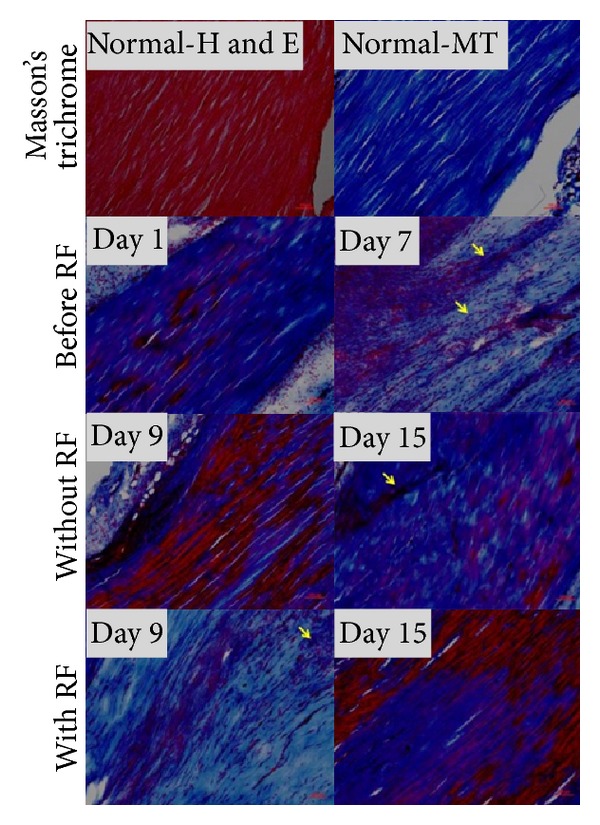
Figure 7.

Masson trichrome (MT) showed increased fibroblasts nuclei (arrows) and collagen which stained black and blue in high cellularity areas (day 1 and 7). On day 9, areas of red stained matrix were reduced in RF-treated tendons when compared with tendon without RF. On day 15, red stained matrix reappeared in tendons with or without RF. Magnification, 100x; scale bar, 100 μm.
