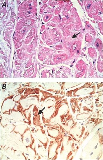
Fig. 2 Photomicrographs show typical findings of cardiac amyloidosis on an endomyocardial biopsy specimen. Arrows indicate the deposition of amyloid. A) Histologic section shows interstitial and perimyocytic deposition of amorphous, light-pink, finely fibrillar eosinophilic material (amyloid) enveloping individual myocytes (H & E, orig. ×400). B) Immunohistochemistry stain confirms the deposition of amyloid (orig. ×400).
