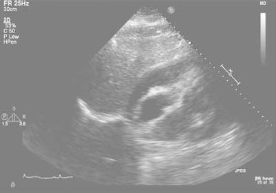
Fig. 4 Echocardiogram reveals a large circumferential pericardial effusion with marked respiratory variation of mitral and tricuspid inflow velocities, plethora of the inferior vena cava, and diastolic compression of the right ventricle, consistent with cardiac tamponade.
