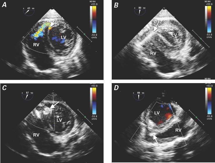
Fig. 4 Patient 6. Transesophageal echocardiograms show a V-shaped apical muscular ventricular septal defect in the right ventricular (RV) inflow region after repair of tetralogy of Fallot. A) Color-flow Doppler mode reveals the defect (arrow) in the posterior portion of the interventricular septum. B) The guidewire (arrow) is passed through the interventricular septum into the left ventricle (LV) by means of a perventricular approach. C) Transgastric short-axis view shows the occluder (arrow), with its left disc not fully opened in the LV side after deployment. D) Midesophageal long-axis view shows the occluder after deployment (arrow).
