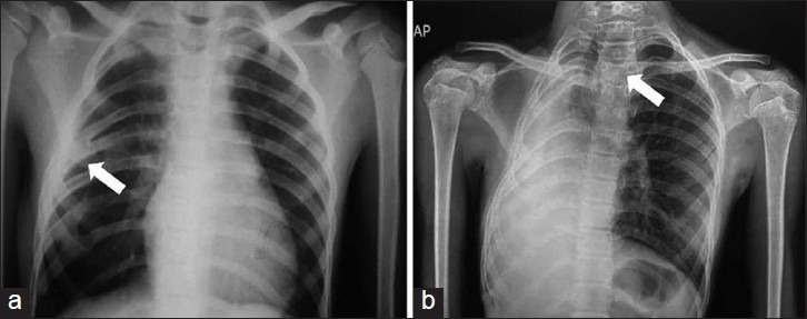
Figure 4.

(a) Rib fusion on X-ray chest (white arrow) (b) X-ray chest showing mediastinal shift, rib crowding, ends of excised rib, and space available for lung (SAL) – 92%. X-ray thoracic spine showing scoliosis with Cobb's angle of 150, with convexity toward left at T3 level (White arrow)
