
Figure 6.
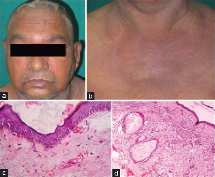
(a) Cutaneous examination of case 3: Heliotrope rash and periorbital edema. (b) Erythema over the “V” area of the neck (i.e., “v” sign) and upper chest. (c) Skin biopsy, 400×, hematoxylin and eosin (H and E): Showing atrophic epidermis, basal cell vacuolar degeneration, dermal edema and perivascular inflammatory infiltrate. (d) Skin biopsy, 400×, H and E: Showing atrophic epidermis, basal vacuolar degeneration, dermal edema and perivascular inflammatory infiltrate with abundant mucin in the dermis