Figure 2.
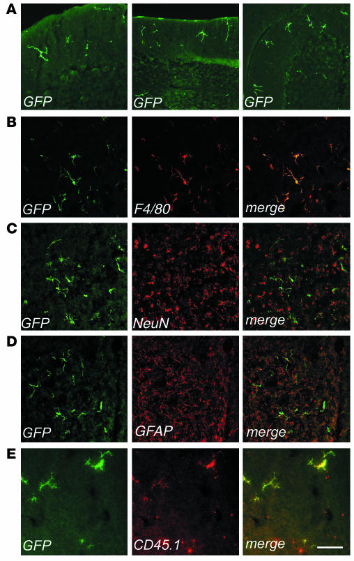
Identification of bone marrow–derived vector-expressing cells in the CNS of transplanted mice. Immunofluorescence analysis of cryostatic sections from the brains of transplanted mice. Fluorescent signals from single optical sections were sequentially acquired and are shown individually and after merging (merge). Immunostaining for GFP (green), F4/80, NeuN, GFAP, or CD45.1 (red) are indicated. (A) Representative sections from the cerebellums of transplanted mice, analyzed at 3 months (left panel, scale bar: 200 μm) and 6 months (middle and right panels, scale bar: 300 μm) after BMT. Three months after BMT, few ramified cells were identified in the upper cortical layers near the meninges. By 6 months after BMT, several GFP+ cells were present in the cortex, and small clusters of ramified GFP+ cells were detected throughout the parenchyma. (B–E) Representative brain sections from transplanted mice 9 months after BMT, immunostained as indicated. (B) GFP+ cells showed a ramified, microglial morphology and F4/80 immunoreactivity. Scale bar: 100 μm. (C and D) Overlay of GFP staining with the neuronal-specific marker NeuN (C) and the astrocytic marker GFAP (D) demonstrated separate localization of the two signals, with GFP+ ramified cells found between neurons and astrocytes. Scale bar: 300 μm. (E) CD45.1 immunostaining identified GFP+ cells as donor-derived. Scale bar: 70 μm.

