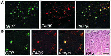
Figure 4.
Enhanced migration and activated morphology of vector-expressing cells in the CNS of As2–/– MLD mice. (A) Cryostatic sections of the corpus callosum of a representative transplanted MLD mouse 6 months after BMT, showing widespread GFP+ cells with a swollen ameboid morphology and coexpressing F4/80. Scale bar: 80 μm. (B) Cryostatic sections from the hippocampus of the same mouse were first immunostained as in A, examined under the fluorescent microscope (three panels on the left) and then stained with PAS (panel on the far right). The GFP+, F4/80+ microglia cells were PAS-reactive, indicating their content of lipid storage granules. Scale bar: 40 μm.

