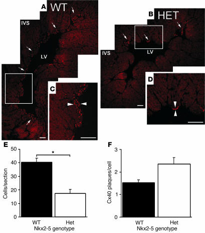
Figure 4.
Cx40 immunohistochemistry confirms hypoplasia of the peripheral Purkinje system and normal cellular expression levels of Cx40 in Nkx2-5+/– mice. Montage confocal images demonstrate Cx40 expression as red, punctate staining in the subendocardial Purkinje fibers of WT (A and C) and Nkx2-5+/– (B and D) left ventricular myocardium. The distribution of Cx40 in the Nkx2-5+/– myocardium is considerably smaller than in WT (arrows in A and B). Higher magnification images of the boxed areas in A and B reveal the increased thickness of Purkinje fiber layers (arrowheads) in the WT (C) compared with Nkx2-5+/– heart (D). LV, left ventricle. Scale bars: 100 μm. (E) Cell counts within Cx40-positive domains reveal that Purkinje cell numbers are reduced by approximately half within sections of Nkx2-5+/– ventricles (P < 0.05). (F) Nkx2-5+/– and WT Purkinje cells contain approximately the same the number of Cx40 particles per cell. Three hearts each from WT and Nkx2-5+/– animals were examined.

