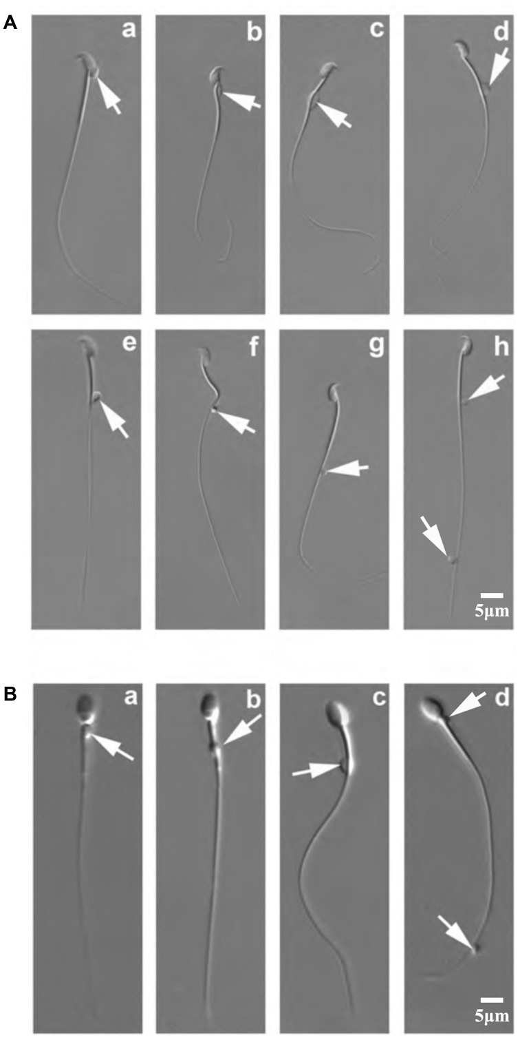
Figure 1.
Positions of CDs on mouse and monkey epididymal spermatozoa. (A) DIC microscopic images showing various positions of CDs on mouse epididymal spermatozoa. Arrows point to CDs, which can be located at the head (a), the neck (b), the middle piece (c), the mid-principal piece junction (d–f), the principal piece (g) and the end piece (h) of the flagellum. (B) DIC microscopic images showing various positions of CDs on monkey epididymal spermatozoa. Arrows point to CDs, which can be located at the neck (a), the middle piece (b), the mid-principal piece junction (c), and the principal/end piece (d) (scale bar=5 µm). CD, cytoplasmic droplet; DIC, differential interference contrast.

