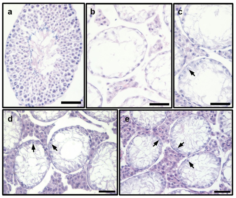
Figure 1.
Morphological observation of the seminiferous tubule in testes of HU-administered mice treated with of 1.5 g kg−1 (LCD+HU), 3.0 g kg−1 (MCD+HU) and 6.0 g kg−1 (HCD+HU) Cistanche deserticola decoctions. (a) normal group, normal cell stage and all types of spermatogenetic cells were observed in the seminiferous epithelium; (b) HU group, severe lumen cavitation of seminiferous tubule in testes was observed, with almost all of the cells degenerated; (c) LCD+HU group; (d) MCD+HU group; (e) HCD+HU group, lumen cavitation of the seminiferous tubule in the testes was observed, with some spermatogonia and early spermatocytes (arrows) present in the seminiferous epithelium. Scale bars=50 µm. CD, Cistanche deserticola; HU, hydroxyurea.

